Differential contribution of Puma and Noxa in dual regulation of p53-mediated apoptotic pathways - PubMed
- ️Sun Jan 01 2006
Differential contribution of Puma and Noxa in dual regulation of p53-mediated apoptotic pathways
Tsukasa Shibue et al. EMBO J. 2006.
Abstract
The activation of tumor suppressor p53 induces apoptosis or cell cycle arrest depending on the state and type of cell, but it is not fully understood how these different responses are regulated. Here, we show that Puma and Noxa, the well-known p53-inducible proapoptotic members of the Bcl-2 family, differentially participate in dual pathways of the induction of apoptosis. In normal cells, Puma but not Noxa induces mitochondrial outer membrane permeabilization (MOMP), and this function is mediated in part by a pathway that involves calcium release from the endoplasmic reticulum (ER) and the subsequent caspase activation. However, upon E1A oncoprotein expression, cells also become susceptible to MOMP induction by Noxa, owing to their sensitization to the ER-independent pathway. These findings offer a new insight into differential cellular responses induced by p53, and may have therapeutic implications in cancer.
Figures

Noxa- and Puma-induced apoptosis in the absence and presence of oncoproteins. (A) Noxa- and Puma-induced apoptosis of NIH3T3 cells expressing various oncoproteins. E1A-3T3, E2F1-3T3 and E7-3T3 cells as well as NIH3T3 cells expressing control vectors (pBabe and pLR) were infected with the retrovirus expressing human (pMx-hNoxa) or mouse Noxa (pMx-mNoxa), human Puma (pMx-hPuma) or the control retrovirus (pMx). Testing with a GFP-expressing pMx construct indicated that the efficiencies of pMx-derived retrovirus infection of control and oncoprotein-expressing NIH3T3 cells were consistently about 80 and 70%, respectively, at 24 h after infection. Values shown are means±s.d. of triplicate samples. (B) Oligomerization of Bax (left panel) and Bak (right panel). Mitochondria-enriched heavy-membrane fractions were prepared from NIH3T3 cells infected with the indicated retrovirus. Obtained protein samples were subsequently treated with the chemical crosslinker 1,6-bismaleimidohexane (BMH) or dimethylsulfoxide (DMSO), and analyzed by immunoblotting. Markers Mo, Di, Tr and Te represent the sizes of monomer, dimer, trimer and tetramer, respectively. (*) An intramolecularly crosslinked Bak monomer. (C, D) MOMP induced by Noxa and Puma in control NIH3T3 cells and E1A-3T3 cells. Membrane insertion of Bax (C) and cytosolic release of cytochrome c (cyt. c) (D) were analyzed. In panel C, cell homogenates were treated with alkali, and subsequently separated by centrifugation. Bax molecules residing in the cytosol or loosely attaching to the membrane are separated into the supernatant (S) fraction, whereas membrane-inserted Bax molecules are separated into the pellet (P) fraction. In panel D, the supernatant and pellet fractions obtained by the digitonin treatment and subsequent centrifugation were analyzed, in which supernatant corresponds to the cytosolic fraction and pellet corresponds to the membrane and nuclear fraction.

zVADfmk-sensitive and -insensitive pathways of MOMP induction. (A) Effect of zVADfmk on Puma- and Noxa-induced cell death in control NIH3T3 cells and E1A-3T3 cells. Under these experimental settings, zVADfmk treatment did not have clear cytotoxic effects. Values shown are means±s.d. from triplicate samples. (B, C) Inhibitory effect of zVADfmk on MOMP. The effects of zVADfmk treatment on membrane insertion of Bax (B) and cytochrome c release (C) were analyzed. In panel C, the amount of cytochrome c detected in the supernatant fractions of Noxa- or Puma- expressing E1A-3T3 cells was reproducibly increased by the zVADfmk treatment. Because the amount of cytochrome c detected in the pellet fraction decreased to a similar extent regardless of zVADfmk treatment, difference in the supernatant fraction is presumably due to the inhibitory effect of zVADfmk on the progression of apoptosis, that is, zVADfmk-treated cells do not readily lose their plasma membrane integrity and thereby they should retain released cytochrome c molecules in the cytosol for longer periods than untreated cells. (D) Puma-induced Bax membrane insertion in wild-type (WT) and Apaf1-deficient (Apaf1−/−) MEFs. WT and Apaf1−/− MEFs were prepared from the same litter. Notably, Puma-induced membrane insertion of Bax occurred in an Apaf-1-independent but caspase-dependent manner.

Essential role of caspase-12 in Puma-induced MOMP and apoptosis. (A) Effects of various peptide-based inhibitors of caspases on Puma-induced Bax membrane insertion. NIH3T3 cells expressing Puma were treated with DMSO, a pan-caspase inhibitor zVADfmk, or various peptide-based inhibitors that specifically inhibit several members of the caspase family (from zWEHDfmk to zLEEDfmk). (B) Effect of zWEHDfmk on Noxa- and Puma-induced cell death in control NIH3T3 cells and E1A-3T3 cells. (C) Caspase-12 (casp-12) processing during Puma-induced apoptosis of NIH3T3 cells and Noxa-induced apoptosis of E1A-3T3 cells. Caspase-12 in the total cell lysate of indicated cells was detected by immunoblotting using the anti-caspase-12 rat monoclonal antibody. The processed form of caspase-12, a possible intermediate for the conversion of the precursor (procasp-12) into the active form, was detected specifically during Puma-induced apoptosis of NIH3T3 cells. Cells were harvested at the indicated times after infection with the pMx-derived retrovirus. (D–F) Effect of shRNA-mediated knock-down of caspase-12 on Puma-induced apoptosis of NIH3T3 cells. NIH3T3 cells were infected with lentivirus that expresses either scramble shRNA or shRNA targeting caspase-12. Three different caspase-12-targeting shRNA sequences (sh casp-12 A–C) were tested, and each resulted in 64, 83 and 63% reduction in the level of procaspase-12 expression, respectively (D). The effects of caspase-12-targeting shRNA on Puma-induced cell death (E) and Bax membrane insertion (F) were also analyzed. In panels B and E, values shown are means±s.d. from triplicate samples.

InsP3R-mediated calcium release from ER during Puma-induced apoptosis. (A, B) Effects of the blockade of ER calcium channels on Puma-induced apoptosis of NIH3T3 cells. Puma-induced cell death (A) and caspase-12 processing (B) in NIH3T3 cells were analyzed in the presence of the blockers of InsP3R (xestC and 2-APB) or dantrolene, the blocker of ryanodine receptor (RyR). (C, D) Impairment of Puma-induced apoptosis by the shRNA-mediated knock-down of type I InsP3R expression. NIH3T3 cells were infected with lentivirus that expresses either scramble shRNA or shRNA targeting type I InsP3R. Three different type I InsP3R-targeting shRNA sequences (sh InsP3RI A–C) were tested, and each resulted in 6, 84 and 60% reduction in the level of type I InsP3R expression, respectively (C). The effects of these shRNA on Puma-induced cell death (D) were also analyzed. (E) Interaction between Bcl-2 family proteins and InsP3Rs. NIH3T3 cells infected with Puma-expressing, Noxa-expressing or control retrovirus were analyzed by immunoprecipitation. The lysate from each sample was immunoprecipitated with an antibody to type I InsP3R (InsP3RI) (upper panel) or an antibody to type III InsP3R (InsP3RIII) (lower panel), and subsequently analyzed by immunoblotting. In panels A and D, values shown are means±s.d. from triplicate samples.

Causal contribution of ER calcium release in Puma-induced apoptosis. (A, B) Time-lapse observation of [Ca2+]c change during Puma-induced apoptosis of control NIH3T3 cells (left panel) and Noxa-induced apoptosis of E1A-3T3 cells (right panel). Cells nucleofected with the YC3.60 expression vector, together with pMx-hPuma or -hNoxa, were analyzed by time-lapse microscopy (see Materials and methods). The time point at which the relevant cell turned PI-positive is set as time 0, and changes in YC3.60 emission ratio (YFP/CFP) were retrospectively displayed. Panel A shows pseudocolored images representing emission ratio calculated on the pixel-by-pixel basis, whereas panel B shows the ratio of fluorescence intensity of CFP and YFP within a region drawn around an individual cell. In panel B, five representative cases are shown from each of control NIH3T3 cells expressing Puma and E1A-3T3 cells expressing Noxa. (C, D) Effects of BAPTA-AM (C) and calpain inhibitor I (calpi-I) (D) on the rate of Puma-induced death of NIH3T3 cells and that of Noxa-induced death of E1A-3T3 cells. Values shown are means±s.d. from triplicate samples. (E) Effects of calpain inhibitor I and BAPTA-AM on Puma-induced caspase-12 processing.

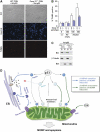
Requirement for caspase-12 in p53-induced apoptosis of CGNs. (A, B) Apoptosis induced by ectopic expression of p53 in wild-type (WT) (+/+) and caspase-12-deficient (casp-12−/−) CGNs. WT and casp-12−/− CGNs were prepared from the same litter and infected with adenovirus expressing p53 (Ad-p53) or control adenovirus; apoptosis was then analyzed by TUNEL. Representative images of CGNs infected with p53-expressing adenovirus at a multiplicity of infection (m.o.i.) of 200 are shown in panel A. Quantitation of TUNEL positivity is shown in panel B, wherein values shown are means±s.d. from four experiments, each using an independent pair of clones. (C) p53-induced Bax membrane insertion in caspase-12-heterozygous (casp-12+/−) and casp-12−/− CGNs. casp-12+/− and casp-12−/− CGNs were prepared from the same litter, and infected with the adenovirus expressing p53 at 200 m.o.i. (D) Dual regulation of mitochondrial apoptotic pathway by p53 targets, Puma and Noxa. In certain types of cells such as normal fibroblasts, thymocytes and CGNs, MOMP is induced at least partly through the zVADfmk-sensitive pathway (left, blue arrow), which is activated by Puma but not Noxa. This zVADfmk-sensitive pathway involves ER events, that is, calcium release through InsP3Rs, which in turn activates calpain. These ER events lead to the activation of caspase-12 and presumably other members of the caspase family; the activation of this pathway, together with the direct inhibition of prosurvival Bcl-2 family proteins by the BH3-only proteins on mitochondria, induces efficient activation of both Bax and Bak, and MOMP induction. Under certain conditions, such as fibroblasts expressing the E1A oncoprotein, a zVADfmk-insensitive MOMP-inducing pathway (right, green arrow), which can be activated by both Puma and Noxa, becomes effective. E1A presumably facilitates the operation of this pathway through the partial activation of Bax. Further studies are required to confirm the validity of this model, particularly regarding the role of Puma in the modulation of InsP3R function, and regarding the role of caspase-12 and other caspases in the activation of Bax and Bak.
Similar articles
-
Nakajima W, Tanaka N. Nakajima W, et al. Biochem Biophys Res Commun. 2011 Oct 7;413(4):643-8. doi: 10.1016/j.bbrc.2011.09.036. Epub 2011 Sep 14. Biochem Biophys Res Commun. 2011. PMID: 21945433
-
Zhang HM, Yuan J, Cheung P, Chau D, Wong BW, McManus BM, Yang D. Zhang HM, et al. Mol Cell Biol. 2005 Jul;25(14):6247-58. doi: 10.1128/MCB.25.14.6247-6258.2005. Mol Cell Biol. 2005. PMID: 15988033 Free PMC article.
-
Mathai JP, Germain M, Marcellus RC, Shore GC. Mathai JP, et al. Oncogene. 2002 Apr 11;21(16):2534-44. doi: 10.1038/sj.onc.1205340. Oncogene. 2002. PMID: 11971188
-
[Lysosomal membrane permeabilization as apoptogenic factor].
Pupyshev AB. Pupyshev AB. Tsitologiia. 2011;53(4):313-24. Tsitologiia. 2011. PMID: 21675210 Review. Russian.
-
Mitochondrial outer membrane permeabilization during apoptosis: the innocent bystander scenario.
Chipuk JE, Bouchier-Hayes L, Green DR. Chipuk JE, et al. Cell Death Differ. 2006 Aug;13(8):1396-402. doi: 10.1038/sj.cdd.4401963. Epub 2006 May 19. Cell Death Differ. 2006. PMID: 16710362 Review.
Cited by
-
Synergistic killing of colorectal cancer cells by oxaliplatin and ABT-737.
Raats DA, de Bruijn MT, Steller EJ, Emmink BL, Borel-Rinkes IH, Kranenburg O. Raats DA, et al. Cell Oncol (Dordr). 2011 Aug;34(4):307-13. doi: 10.1007/s13402-011-0026-8. Epub 2011 Apr 6. Cell Oncol (Dordr). 2011. PMID: 21468686
-
Moncunill-Massaguer C, Saura-Esteller J, Pérez-Perarnau A, Palmeri CM, Núñez-Vázquez S, Cosialls AM, González-Gironès DM, Pomares H, Korwitz A, Preciado S, Albericio F, Lavilla R, Pons G, Langer T, Iglesias-Serret D, Gil J. Moncunill-Massaguer C, et al. Oncotarget. 2015 Dec 8;6(39):41750-65. doi: 10.18632/oncotarget.6154. Oncotarget. 2015. PMID: 26497683 Free PMC article.
-
Genome-wide high-resolution aCGH analysis of gestational choriocarcinomas.
Poaty H, Coullin P, Peko JF, Dessen P, Diatta AL, Valent A, Leguern E, Prévot S, Gombé-Mbalawa C, Candelier JJ, Picard JY, Bernheim A. Poaty H, et al. PLoS One. 2012;7(1):e29426. doi: 10.1371/journal.pone.0029426. Epub 2012 Jan 9. PLoS One. 2012. PMID: 22253721 Free PMC article.
-
Orphan receptor NR4A3 is a novel target of p53 that contributes to apoptosis.
Fedorova O, Petukhov A, Daks A, Shuvalov O, Leonova T, Vasileva E, Aksenov N, Melino G, Barlev NA. Fedorova O, et al. Oncogene. 2019 Mar;38(12):2108-2122. doi: 10.1038/s41388-018-0566-8. Epub 2018 Nov 19. Oncogene. 2019. PMID: 30455429
-
Identification of New Molecular Biomarkers in Ovarian Cancer Using the Gene Expression Profile.
Olbromski PJ, Pawlik P, Bogacz A, Sajdak S. Olbromski PJ, et al. J Clin Med. 2022 Jul 4;11(13):3888. doi: 10.3390/jcm11133888. J Clin Med. 2022. PMID: 35807169 Free PMC article.
References
-
- Blomgren K, Zhu C, Wang X, Karlsson JO, Leverin AL, Bahr BA, Mallard C, Hagberg H (2001) Synergistic activation of caspase-3 by m-calpain after neonatal hypoxia–ischemia: a mechanism of ‘pathological apoptosis'? J Biol Chem 276: 10191–10198 - PubMed
-
- Boehning D, Patterson RL, Sedaghat L, Glebova NO, Kurosaki T, Snyder SH (2003) Cytochrome c binds to inositol (1,4,5) trisphosphate receptors, amplifying calcium-dependent apoptosis. Nat Cell Biol 5: 1051–1061 - PubMed
-
- Breckenridge DG, Germain M, Mathai JP, Nguyen M, Shore GC (2003) Regulation of apoptosis by endoplasmic reticulum pathways. Oncogene 22: 8608–8618 - PubMed
-
- Brown JM, Attardi LD (2005) The role of apoptosis in cancer development and treatment response. Nat Rev Cancer 5: 231–237 - PubMed
-
- Chen L, Willis SN, Wei A, Smith BJ, Fletcher JI, Hinds MG, Colman PM, Day CL, Adams JM, Huang DC (2005) Differential targeting of prosurvival Bcl-2 proteins by their BH3-only ligands allows complementary apoptotic function. Mol Cell 17: 393–403 - PubMed
Publication types
MeSH terms
Substances
LinkOut - more resources
Full Text Sources
Other Literature Sources
Molecular Biology Databases
Research Materials
Miscellaneous