High-content analysis of antibody phage-display library selection outputs identifies tumor selective macropinocytosis-dependent rapidly internalizing antibodies - PubMed
High-content analysis of antibody phage-display library selection outputs identifies tumor selective macropinocytosis-dependent rapidly internalizing antibodies
Kevin D Ha et al. Mol Cell Proteomics. 2014 Dec.
Abstract
Many forms of antibody-based targeted therapeutics, including antibody drug conjugates, utilize the internalizing function of the targeting antibody to gain intracellular entry into tumor cells. Ideal antibodies for developing such therapeutics should be capable of both tumor-selective binding and efficient endocytosis. The macropinocytosis pathway is capable of both rapid and bulk endocytosis, and recent studies have demonstrated that it is selectively up-regulated by cancer cells. We hypothesize that receptor-dependent macropinocytosis can be achieved using tumor-targeting antibodies that internalize via the macropinocytosis pathway, improving potency and selectivity of the antibody-based targeted therapeutic. Although phage antibody display libraries have been utilized to find antibodies that bind and internalize to target cells, no methods have been described to screen for antibodies that internalize specifically via macropinocytosis. We hereby describe a novel screening strategy to identify phage antibodies that bind and rapidly enter tumor cells via macropinocytosis. We utilized an automated microscopic imaging-based, High Content Analysis platform to identify novel internalizing phage antibodies that colocalize with macropinocytic markers from antibody libraries that we have generated previously by laser capture microdissection-based selection, which are enriched for internalizing antibodies binding to tumor cells in situ residing in their tissue microenvironment (Ruan, W., Sassoon, A., An, F., Simko, J. P., and Liu, B. (2006) Identification of clinically significant tumor antigens by selecting phage antibody library on tumor cells in situ using laser capture microdissection. Mol. Cell. Proteomics. 5, 2364-2373). Full-length human IgG molecules derived from macropinocytosing phage antibodies retained the ability to internalize via macropinocytosis, validating our screening strategy. The target antigen for a cross-species binding antibody with a highly active macropinocytosis activity was identified as ephrin type-A receptor 2. Antibody-toxin conjugates created using this macropinocytosing IgG were capable of potent and receptor-dependent killing of a panel of EphA2-positive tumor cell lines in vitro. These studies identify novel methods to screen for and validate antibodies capable of receptor-dependent macropinocytosis, allowing further exploration of this highly efficient and tumor-selective internalization pathway for targeted therapy development.
© 2014 by The American Society for Biochemistry and Molecular Biology, Inc.
Figures

Outline of screening strategy and data from the first step of the screening, i.e. phage binding to DU145 cells. A, Schematic of HCA screening to identify macropinocytosis-dependent antibodies. HCA instruments allow automated high throughput detection of antibody colocalization with a macropinocytosis marker. The starting materials for the screening are sublibraries generated previously by us from LCM-based phage antibody library selection (1) that are enriched for internalizing phage antibodies binding to tumor cells in situ. B, DU145 cells were incubated in 96-well plates with phage-containing supernatants for 24 h at 37 °C in complete DMEM and10% FBS. Nuclei were stained with Hoechst 33342. Bound phages were immunolabeled with anti-fd antibodies (green). Zoomed insert portrays software-based, automated cell analysis, measuring mean fluorescence intensities (MFI) of immunolabeled phages. Over 300 cells were quantified for each phage clone. C, Plot of MFI values of immunolabeled phage binding to cell for 1439 phage clones. Red horizontal line represents MFI of ∼250,000, the threshold for prioritizing clones for further internalization analysis.

Colocalization of phage antibodies with the macropinocytosis marker ND70-TR. A, Epifluorescent images of DU145 cells that were incubated with phage-containing supernatants and 50 μg/ml ND70-TR (red) for 24 h at 37 °C. Cell-associated phage were then detected by biotin-labeled anti-fd antibody followed by streptavidin-AlexaFluor 488 (green). Colocalization results in color overlap (yellow). Scale bar denotes 20 μm. B, To analyze colocalization, arbitrary lines were drawn across cells and fluorescent intensities along the drawn line were plotted for phages (green) and ND70-TR fluorescence (red). Co-variation of line intensity indicates colocalization. Representative images of two different phage antibodies with differing colocalization patterns are shown. C, Pearson's correlation coefficient (PCC) was quantified and averaged from >30 cells per phage conditions. Error bars denote S.E. for n = 3; * and ** indicate p values of <0.05 and <0.01, respectively, using two-tailed student's T-tests assuming unequal variance. D, Colocalization screening. DU145 cells were plated onto 96-well plates and incubated with phages and ND70-TR (red) for 24 h at 37 °C. Cells were immunolabeled against bacteriophages (green) and nuclei were stained with Hoechst 33342 (blue). E, Mean PCC between immunolabeled phage and ND70-TR of 360 phage clones, quantified from minimum of 300 cells per phage clone. PCC values were normalized to control phage clones that exhibited poor internalization. Green horizontal line represents 200% of control, a threshold for further analysis.

Confocal analysis of phage antibody internalization by DU145 cells. Confocal Z-slices of DU145 cells incubated with purified phage for A, 24 h at 37 °C or B, 8 h at 37 °C in the presence of ND70-TR. Cells were immunolabeled against phages (green), lysosomes (LAMP1, red), and nuclei (Hoechst 33342, blue). Scale bar: 20 μm. C, Mean PCC of internalized phages and ND70-TR. Over 30 cells were analyzed per phage antibody. ** denotes two-tailed t test p values of <0.01. Error bars represent S.E. for n = 3.

Internalization and colocalization analysis of IgGs derived from scFvs. A, DU145 cells co-incubated with three IgGs with different internalization properties at 10 μg/ml and 50 μg/ml ND70-TR (red) for 90 min at 37 °C. Cells were immunolabeled against IgG using anti-human Fc (green). Nuclei were stained with Hoechst 33342 (blue). Single confocal Z-slice images are shown. Scale bar: 20 μm. B, PCC analysis of colocalization of IgGs HCA-F1, HCA-M1, and HCA-S1 with ND70-TR using Z-slices crossing the entire cell, quantitating a minimum of 10 cells. ** and *** denote two-tailed t test p values of <0.01 and <0.001, respectively. Error bars represent S.E. for n = 3.

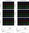
Kinetics of antibody internalization and subcellular localization. DU145 cells were incubated with three different IgGs (HCA-F1, HCA-M1, or HCA-S1) at 10 μg/ml for 15 min at 4 °C and then chased with complete DMEM/10% FBS for indicated time periods. Cells were then fixed, permeabilized, and immunolabeled against human IgG (green) and A, early endosomes (EEA1, red) or B, lysosomes (LAMP1, red). Nuclei were stained with Hoechst 33342 (blue). Scale bar: 20 μm. Pearson's correlation coefficients between immunolabeled C, EEA1 or D, LAMP1 and immunolabeled IgG were averaged from a minimum of 30 cells. Error bars denote S.E. of n = 3.

Macropinocytosis inhibitors prevent internalization of IgG HCA-F1. DU145 cells were pretreated with 50 μg/ml cytochalasin D, 7.5 μg/ml EIPA, or DMSO (control) for 30 min at 37 °C followed by co-incubation with 10 μg/ml IgG HCA-F1 and ND70-TR (red) in the presence of cytochalasin D, EIPA, or DMSO in complete DMEM/10% FBS for 40 min at 37 °C. Cells were then immunolabeled for human IgG (green). Nuclei were stained with Hoechst 33342 (blue). A, Individual confocal Z-slices of representative cells. CytoD: cytochalasin D. Scale bar: 20 μm. B, The percentage of internalized IgG HCA-F1 was quantitated by measuring the ratio of internalized, cytosolic IgG HCA-F1 fluorescence over total cell IgG HCA-F1 fluorescence, analyzing >15 cells over three independent experiments. *** indicates p value of <0.001 using two-tailed student's t test assuming unequal variance. Error bars represent S.E. with n = 3.

EphA2 identified as target antigen bound by macropinocytosing antibody IgG HCA-F1. A, Immunoprecipiation of the target antigen from surface-biotinylated Du145 whole cell lysates using scFv HCA-F1-Fc fusion immobilized onto a solid matrix. The immunoprecipitation product was run on SDS-PAGE and subjected to Western blot analysis using streptavidin-HRP to locate the position of membrane proteins. The dominant band, denoted by “*,” represents the approximate region from which the corresponding SDS-PAGE gel was extracted for mass spectrometry analysis. B, Binding to ectopically expressed EphA2. Chinese hamster ovarian (CHO) cells were co-transfected with pEGFP-N2 (to label transfected cells) and pCMV6 expression constructs bearing either human EphA2 or Lgr5 (control). Cells were then incubated with IgG HCA-F1, followed by immunolabeling using anti-human Fc AlexaFluor® 647. Cells were gated for GFP expression and plotted for AlexaFluor® 647 fluorescence (FL4). C, Plot of MFI values as analyzed by FACS. IgG HCA-F1 binds specifically to ectopically expressed EphA2, confirming the target identification.

Functional internalization assay using IgG HCA-F1-toxin conjugates. A, FACS analysis showing EphA2-positive (DU145) and EphA2-negative (LNCaP, control) cells. IgG HCA-F1 was incubated with the cells and binding was detected with anti-human Fc. MFI values are shown in the far right panel. B, IgG HCA-F1 was conjugated to saporin and incubated with target (DU145) and control (LNCaP) cells. Controls: toxin only and IgG HCA-F1 only. Cell viability was measured 4 days later using the CCK-8 assay.
Similar articles
-
Ha KD, Bidlingmaier SM, Su Y, Lee NK, Liu B. Ha KD, et al. Methods Enzymol. 2017;585:91-110. doi: 10.1016/bs.mie.2016.10.004. Epub 2016 Dec 1. Methods Enzymol. 2017. PMID: 28109445 Free PMC article.
-
Discovery of internalizing antibodies to tumor antigens from phage libraries.
Zhou Y, Marks JD. Zhou Y, et al. Methods Enzymol. 2012;502:43-66. doi: 10.1016/B978-0-12-416039-2.00003-3. Methods Enzymol. 2012. PMID: 22208981 Free PMC article.
-
Immunotoxins and other conjugates containing saporin-s6 for cancer therapy.
Polito L, Bortolotti M, Pedrazzi M, Bolognesi A. Polito L, et al. Toxins (Basel). 2011 Jun;3(6):697-720. doi: 10.3390/toxins3060697. Epub 2011 Jun 22. Toxins (Basel). 2011. PMID: 22069735 Free PMC article. Review.
-
Ferrini S, Sforzini S, Canevari S. Ferrini S, et al. Methods Mol Biol. 2001;166:177-92. doi: 10.1385/1-59259-114-0:177. Methods Mol Biol. 2001. PMID: 11217367 Review. No abstract available.
Cited by
-
Bidlingmaier S, Su Y, Liu B. Bidlingmaier S, et al. Methods Mol Biol. 2015;1319:51-63. doi: 10.1007/978-1-4939-2748-7_3. Methods Mol Biol. 2015. PMID: 26060069 Free PMC article.
-
Li C, Wang Y, Liu T, Niklasch M, Qiao K, Durand S, Chen L, Liang M, Baumert TF, Tong S, Nassal M, Wen YM, Wang YX. Li C, et al. Antiviral Res. 2019 Feb;162:118-129. doi: 10.1016/j.antiviral.2018.12.019. Epub 2018 Dec 30. Antiviral Res. 2019. PMID: 30599174 Free PMC article.
-
Intracellular nanoparticle delivery by oncogenic KRAS-mediated macropinocytosis.
Liu X, Ghosh D. Liu X, et al. Int J Nanomedicine. 2019 Aug 16;14:6589-6600. doi: 10.2147/IJN.S212861. eCollection 2019. Int J Nanomedicine. 2019. PMID: 31496700 Free PMC article.
-
High Content Imaging (HCI) on Miniaturized Three-Dimensional (3D) Cell Cultures.
Joshi P, Lee MY. Joshi P, et al. Biosensors (Basel). 2015 Dec 14;5(4):768-90. doi: 10.3390/bios5040768. Biosensors (Basel). 2015. PMID: 26694477 Free PMC article. Review.
-
ALPPL2 Is a Highly Specific and Targetable Tumor Cell Surface Antigen.
Su Y, Zhang X, Bidlingmaier S, Behrens CR, Lee NK, Liu B. Su Y, et al. Cancer Res. 2020 Oct 15;80(20):4552-4564. doi: 10.1158/0008-5472.CAN-20-1418. Epub 2020 Aug 31. Cancer Res. 2020. PMID: 32868383 Free PMC article.
References
-
- Ruan W., Sassoon A., An F., Simko J. P., Liu B. (2006) Identification of clinically significant tumor antigens by selecting phage antibody library on tumor cells in situ using laser capture microdissection. Mol. Cell. Proteomics. 5, 2364–2373 - PubMed
-
- Burris H. A., 3rd, Tibbitts J., Holden S. N., Sliwkowski M. X., Lewis Phillips G. D. (2011) Trastuzumab emtansine (T-DM1): a novel agent for targeting HER2+ breast cancer. Clin. Breast Cancer 11, 275–282 - PubMed
-
- Sievers E. L., Senter P. D. (2013) Antibody-drug conjugates in cancer therapy. Annu. Rev. Med. 64, 15–29 - PubMed
Publication types
MeSH terms
Substances
LinkOut - more resources
Full Text Sources
Other Literature Sources
Miscellaneous