Overactive type 2 cannabinoid receptor induces meiosis in fetal gonads and impairs ovarian reserve - PubMed
- ️Sun Jan 01 2017
Overactive type 2 cannabinoid receptor induces meiosis in fetal gonads and impairs ovarian reserve
Emanuela De Domenico et al. Cell Death Dis. 2017.
Abstract
Type 2 cannabinoid receptor (CB2R) has been proposed to promote in vitro meiotic entry of postnatal male germ cells and to maintain the temporal progression of spermatogenesis in vivo. However, no information is presently available on the role played by CB2R in male and female fetal gonads. Here we show that in vitro pharmacological stimulation with JWH133, a CB2R agonist, induced activation of the meiotic program in both male and female fetal gonads. Upon stimulation, gonocytes initiated the meiotic program but became arrested at early stages of prophase I, while oocytes showed an increased rate of meiotic entry and progression toward more advanced stage of meiosis. Acceleration of meiosis in oocytes was accompanied by a strong increase in the percentage of γ-H2AX-positive pachytene and diplotene cells, paralleled by an increase of TUNEL-positive cells, suggesting that DNA double-strand breaks were not correctly repaired during meiosis, leading to oocyte apoptosis. Interestingly, in vivo pharmacological stimulation of CB2R in fetal germ cells through JWH133 administration to pregnant females caused a significant reduction of primordial and primary follicles in the ovaries of newborns with a consequent depletion of ovarian reserve and reduced fertility in adult life, while no alterations of spermatogenesis in the testis of the offspring were detected. Altogether our findings highlight a pro-meiotic role of CB2R in male and female germ cells and suggest that the use of cannabis in pregnant female might represent a risk for fertility and reproductive lifespan in female offspring.
Conflict of interest statement
The authors declare no conflict of interest.
Figures
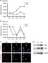
Cannabinoid receptors CB1 and CB2 are expressed in fetal male and female gonads. (a) Relative expression (2−ΔCt) of CB1R and CB2R in male fetal gonad and (b) in female fetal gonad at different times of development. (c) Immunofluorescence analysis for CB2R in both E15.5 male and female fetal germ cells shows the membrane localization of the receptor. (d) Western blotting analysis of CB2R in isolated E15.5 female (F) and male (M) fetal germ cells. Isolated 7 dpn spermatogonia (SPG) were used as positive control. VASA is used as a marker of germ cells

Activation of CB2R promotes gonocyte meiotic entry. (a) Representative immunofluorescence images showing SCP3 (green) organization on nuclear spreads at the stages of preleptotene, early leptotene and leptotene cells of meiotic prophase I. (b) Histogram representing the percentage of nuclei with meiotic SCP3 staining in male germ cells from E13.5 gonads after 48 h of culture in the absence or presence of JWH133 alone or in combination with AM630. (c) Percentage of preleptotene, early leptotene and leptotene nuclei in cultures of E13.5 male germ cells treated and untreated for 48 h with JWH133 alone or in combination with AM630. (d) Histogram representing the percentage of nuclei with meiotic SCP3 staining in E15.5 male germ cells treated and untreated for 48 h with JWH133 alone or in combination with AM630. (e) Percentage of preleptotene, early leptotene and leptotene nuclei in cultures of E15.5 male germ cells treated and untreated for 48 h with JWH133 alone or in combination with AM630. (f and g) Real-time PCR of meiotic genes Stra8 and Nanos2 in E13.5 male germ cells treated or not for 48 h with JWH133. Data were collected from at least three different experiments, using a minimum of 10 embryos for each one. *P<0.05 and **P<0.01
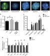
Activation of CB2R promotes fetal oocyte meiotic entry at E13.5. (a) Representative immunofluorescence images showing SCP3 (green) organization on nuclear spreads at the stages of preleptotene, early leptotene, leptotene, zygotene and pachytene cells of meiotic prophase I. (b) Histogram representing the percentage of nuclei with meiotic SCP3 staining in female germ cells from E13.5 gonads treated or not with JWH133 alone or in combination with AM630 for 48 h. (c) Percentage of meiotic nuclei at different stages of prophase I in female germ cells from E13.5 gonads treated or not for 48 h with JWH133 alone or in combination with AM630. (d) Real-time PCR of meiotic genes Dmc1, Spo11, c-Kit, SCP1, SCP3, Stra8 and Nanos2 in female germ cells from E13.5 gonads treated or not for 48 h with JWH133. Data were collected from at least three different experiments, using a minimum of 10 embryos for each one. *P<0.05 and **P<0.01

Activation of CB2R promotes fetal oocyte meiotic progression at E15.5. (a) Representative immunofluorescence images showing SCP3 (green) organization on nuclear spreads at the stages of leptotene, zygotene, pachytene, diplotene and metaphase-like cells of meiotic prophase I. (b) Histogram representing the percentage of nuclei with meiotic SCP3 staining in female germ cells from E15.5 gonads treated or not with JWH133 for 48 h. (c) Percentage of meiotic nuclei at different stages of prophase I of female germ cells from E15.5 gonads treated or not for 48 h with JWH133. Data were collected from at least three different experiments, using a minimum of 10 embryos for each one. NS, not significant. *P<0.05
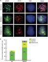
Activation of CB2R increases γ-H2AX foci in fetal oocytes. (a) Representative immunofluorescence images showing staining of SCP3 (red) and γ-H2AX (green) on pachytene, diplotene and metaphase-like cells from E15.5 ovary. (b) Percentage of double-positive SCP3 and γ-H2AX oocytes at the stages of pachytene, diplotene and metaphase-like from E15.5 ovary, treated or not with JWH133 for 48 h. Data were collected from at least three different experiments, using a minimum of 10 embryos for each one
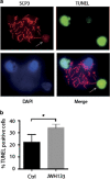
Activation of CB2R increases the number of terminal deoxinucleotidyl transferase-mediated dUTP-fluorescein nick end labeling (TUNEL)-positive oocytes. (a) Representative image of spreads nuclei of E15.5 female germ cells stained for SCP3 (red) and TUNEL (green). An SCP3-positive cell at the pachytene stage, a TUNEL-positive cell with disassembled pattern of synaptonemal complex (indicated by white arrow) and TUNEL-negative cells are shown. (b) Percentage of SCP3 and TUNEL double positive in female germ cells from E15.5 gonads treated or not with JWH133 for 48 h. Data were collected from at least three different experiments, using a minimum of 10 embryos for each one. *P<0.05
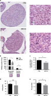
In vivo treatment of pregnant mice with JWH133 affect ovarian reserve of female pups. (a) Morphological staining with H&E of 1 dpn ovaries in utero exposed to JWH133 shows a reduction in follicle number and size as shown at higher magnification on the right (Pr.: primordial follicles). (b) The histograms show a significant reduction in primordial and primary follicle number in the ovary of F1 offspring in utero exposed to JWH133 with respect to vehicle-exposed offspring. (c) A significant reduction in the diameter of primordial follicles was detected in the ovaries of F1 from JWH133-treated pregnant mice with respect to F1 primordial follicles from control pregnant mice. (d) Mating rate of F1 female from JWH133-treated pregnant mice was unchanged with respect to mating rate of F1 control mice. (e) Litter size of F2 from JWH133 F1 female was significantly reduced with respect to that F1 control mice. Data were collected using a minimum of three 1 dpn female pups. NS, not significant. *P<0.05

Schematic representation of the effects of CB2R overactivation on fetal ovary. Pregnant females were intraperitoneally injected with JWH133 for 4 consecutive days at the onset of meiosis in oocytes (E12.5). Morphological analysis of F1 ovary at birth (1 dpn) showed a reduced number of primordial (Pr) and primary (Pry) follicles with respect to control. F1 JWH133 female crossed with untreated male showed a decreased fertility with a smaller litter size at F2
Similar articles
-
Dutta S, Burks DM, Pepling ME. Dutta S, et al. Reprod Biol Endocrinol. 2016 Dec 5;14(1):82. doi: 10.1186/s12958-016-0218-1. Reprod Biol Endocrinol. 2016. PMID: 27919266 Free PMC article.
-
Type 2 cannabinoid receptor contributes to the physiological regulation of spermatogenesis.
Di Giacomo D, De Domenico E, Sette C, Geremia R, Grimaldi P. Di Giacomo D, et al. FASEB J. 2016 Apr;30(4):1453-63. doi: 10.1096/fj.15-279034. Epub 2015 Dec 15. FASEB J. 2016. PMID: 26671998
-
Dumont L, Rives-Feraille A, Delessard M, Saulnier J, Rondanino C, Rives N. Dumont L, et al. Andrology. 2021 Mar;9(2):673-688. doi: 10.1111/andr.12928. Epub 2020 Nov 16. Andrology. 2021. PMID: 33112479
-
The developmental origins of the mammalian ovarian reserve.
Grive KJ, Freiman RN. Grive KJ, et al. Development. 2015 Aug 1;142(15):2554-63. doi: 10.1242/dev.125211. Development. 2015. PMID: 26243868 Free PMC article. Review.
-
De Felici M, Klinger FG, Farini D, Scaldaferri ML, Iona S, Lobascio M. De Felici M, et al. Reprod Biomed Online. 2005 Feb;10(2):182-91. doi: 10.1016/s1472-6483(10)60939-x. Reprod Biomed Online. 2005. PMID: 15823221 Review.
Cited by
-
Martínez-Peña AA, Lee K, Pereira M, Ayyash A, Petrik JJ, Hardy DB, Holloway AC. Martínez-Peña AA, et al. Int J Mol Sci. 2022 Jul 20;23(14):8000. doi: 10.3390/ijms23148000. Int J Mol Sci. 2022. PMID: 35887347 Free PMC article.
-
Cannabinoid Receptors Signaling in the Development, Epigenetics, and Tumours of Male Germ Cells.
Barchi M, Innocenzi E, Giannattasio T, Dolci S, Rossi P, Grimaldi P. Barchi M, et al. Int J Mol Sci. 2019 Dec 18;21(1):25. doi: 10.3390/ijms21010025. Int J Mol Sci. 2019. PMID: 31861494 Free PMC article. Review.
-
The Impact of Early Life Exposure to Cannabis: The Role of the Endocannabinoid System.
Martínez-Peña AA, Perono GA, Gritis SA, Sharma R, Selvakumar S, Walker OS, Gurm H, Holloway AC, Raha S. Martínez-Peña AA, et al. Int J Mol Sci. 2021 Aug 9;22(16):8576. doi: 10.3390/ijms22168576. Int J Mol Sci. 2021. PMID: 34445282 Free PMC article. Review.
-
Cannabis alters epigenetic integrity and endocannabinoid signalling in the human follicular niche.
Fuchs Weizman N, Wyse BA, Szaraz P, Defer M, Jahangiri S, Librach CL. Fuchs Weizman N, et al. Hum Reprod. 2021 Jun 18;36(7):1922-1931. doi: 10.1093/humrep/deab104. Hum Reprod. 2021. PMID: 33954787 Free PMC article.
-
Rossi G, Di Nisio V, Chiominto A, Cecconi S, Maccarrone M. Rossi G, et al. Int J Mol Sci. 2023 Apr 19;24(8):7542. doi: 10.3390/ijms24087542. Int J Mol Sci. 2023. PMID: 37108704 Free PMC article.
References
-
- Hilscher B, Hilscher W, Bulthoff-Ohnolz B, Kramer U, Birke A, Pelzer H et al. Kinetics of gametogenesis. Comparative histological and autoradiographic studies of oocytes and transitional prospermatogonia during oogenesis and prespermatogenesis. Cell Tissue Res 1974; 154: 443–470. - PubMed
-
- Western PS, Miles DC, van den Bergen JA, Burton M, Sinclair AH. Dynamic regulation of mitotic arrest in fetal male germ cells. Stem Cells 2008; 26: 339–347. - PubMed
-
- Bowles J, Knight D, Smith C, Wilhelm D, Richman J, Mamiya S et al. Retinoid signaling determines germ cell fate in mice. Science 2006; 312: 596–600. - PubMed
-
- Pellegrini M, Filipponi D, Gori M, Barrios F, Lolicato F, Grimaldi P et al. ATRA and KL promote differentiation toward the meiotic program of male germ cells. Cell Cycle 2008; 7: 3878–3888. - PubMed
MeSH terms
Substances
LinkOut - more resources
Full Text Sources
Other Literature Sources