The Cambrian cirratuliform Iotuba denotes an early annelid radiation - PubMed
- ️Sun Jan 01 2023
The Cambrian cirratuliform Iotuba denotes an early annelid radiation
ZhiFei Zhang et al. Proc Biol Sci. 2023.
Abstract
The principal animal lineages (phyla) diverged in the Cambrian, but most diversity at lower taxonomic ranks arose more gradually over the subsequent 500 Myr. Annelid worms seem to exemplify this pattern, based on molecular analyses and the fossil record: Cambrian Burgess Shale-type deposits host a single, early-diverging crown-group annelid alongside a morphologically and taxonomically conservative stem group; the polychaete sub-classes diverge in the Ordovician; and many orders and families are first documented in Carboniferous Lagerstätten. Fifteen new fossils of the 'phoronid' Iotuba (=Eophoronis) chengjiangensis from the early Cambrian Chengjiang Lagerstätte challenge this picture. A chaetal cephalic cage surrounds a retractile head with branchial plates, affiliating Iotuba with the derived polychaete families 'Flabelligeridae' and Acrocirridae. Unless this similarity represents profound convergent evolution, this relationship would pull back the origin of the nested crown groups of Cirratuliformia, Sedentaria and Pleistoannelida by tens of millions of years-indicating a dramatic unseen origin of modern annelid diversity in the heat of the Cambrian 'explosion'.
Keywords: Cambrian explosion; annelid evolution; body plans; phylogenetics; polychaetes.
Conflict of interest statement
We declare we have no competing interests.
Figures

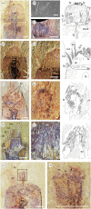
Iotuba chengjiangensis. (a–c) ELI-S-001, complete specimen with recurved gut and head partly retracted; (b) normalized elemental abundance measured by micro-X-ray fluorescence: blue channel, Al + K; green, Si + Zn + N; red, P + Fe; (d–f) ELI-S-002A, anterior trunk with boudinaged gut; head preserved perpendicular to plane of splitting; blue channel in (f): normalized abundance of Al + K; green, Si; red, P + Fe + Cr + Cu; (g) ELI-S-007A, anterior end everted; chancelloriid associated with posterior trunk; the iron-rich region anterior to the head is on a different surface and is not part of the Iotuba fossil; (h) ELI-S-003B, chancelloriid associated with posterior trunk. High-resolution images at Figshare [32]. Scale bars: 10 mm except enlargements (c,e–f), 2 mm. Abbreviations: ch, chancelloriid; con, constriction between boudins; fa, fascicle of spines; fg, foregut; gr, transverse groove; hd, everted head; lt, lateral tube; mg, midgut.

Iotuba chengjiangensis head. (a–c) ELI-S-007AB, branchiae represented in black in interpretative drawings; (b) backscatter scanning electron micrograph showing relief and elevated iron content of branchial filaments; (c) flipped image of counterpart corresponding to region boxed in (a) showing palisade of spines; (d,e) ELI-S-010, head partly retracted, flanked by palisade and at least four fascicles of spines, with distal branchiae; (f,g) ELI-S-003B; head partially retracted; branchiae visible beneath palisades; (h,i) ELI-S-011, head partially retracted, showing branchiae and fascicles of spines; (j,k) ELI-S-004A, partially withdrawn head showing longitudinal (left arrow) and transverse (right arrow) orientation of branchial filaments, which remain terminal even as head is withdrawn. High-resolution images at Figshare [32]. Scale bars: (a,c–f,h,j–k), 2 mm; (b,g,i), 200 µm. Abbreviations: be, basal element of palisade; br, branchiae; fa, fascicle of spines; fg, foregut; pa, palisade of spines.

Internal anatomy of Iotuba chengjiangensis. (a,b) ELI-S-006, midgut and lateral mineral-filled tubes; (c) ELI-S-004A, distinct preservation of foregut; (d) ELI-S-007A, lateral mineral-filled tubes parallel to midgut; (e,f) ELI-S-009, posterior trunk, showing distal termination of lateral tubes; (g–i) ELI-S-005A; (h) anterior termination of lateral tubes; (i) (counterpart, image flipped), coarse mineral grains in gut; (j,k) ELI-S-008, folded specimen with bulb-shaped foregut. High-resolution images at Figshare [32]. Scale bars: 10 mm except enlargements (b,d,f,h–i,k,n), 1 mm. Abbreviations: fg, foregut; lt, lateral tube; mg, midgut.

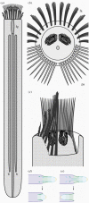
Reconstruction of Iotuba. (a) Dorsal view; (b) anterior view; (c), dorsal view, right-hand fascicles omitted to display retracted head; (d,e), schematic of a hypothetical worm showing withdrawal of an eversible head by: (d), retraction, as in Iotuba; (e), involution, as in ecdysozoan worms. Abbreviations: fa, fascicle of spines; fg, foregut; lt, lateral tube; pa, palisade of spines.

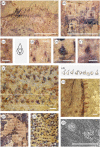
Epidermal ornament in Iotuba chengjiangensis. (a–f) ELI-S-007A, conical, anterior-directed papillae on head; outline of circular base prominent in (b,c,e,f); basal invagination visible in (c,d); (g) ELI-S-002A, detailed outline of trunk papillae; (h) reconstruction of original trunk papilla morphology, corresponding to boxed region in (g); (i), ELI-S-004A, outline of trunk papillae preserved on lateral margin of trunk; (j–k) ELI-S-011, impressions of papillae on inner (j) and outer (k) surfaces of trunk; (l), ELI-S-005A, electron micrograph showing pyrite pseudomorphs in papilla cavities. High-resolution images at Figshare [32]. Scale bars: 200 µm, except (a,b) (2 mm).

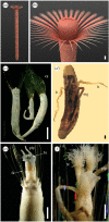
Comparison of Iotuba with extant flabelligerids: (a,b) life reconstruction of Iotuba; (c–f) photographs of extant flabelligerids by Sergio Salazar-Vallejo, reproduced with permission from the copyright holders (withheld from open access agreement): (c) Semiodera tenera [35], with well-displayed cephalic cages, heads partly or fully retracted; (d) dissection of Brada inhabilis [36], showing extensive nephridia; (e), Stylaroides monilifer [37], everted head showing palps and branchial filaments; (f) Stylaroides hirsutus [37], pair of fully everted branchiae. (e,f) Copyright © Unione Zoologica Italiana, reprinted by permission of Taylor & Francis Ltd,
http://www.tandfonline.comon behalf of Unione Zoologica Italiana. Scale bars: 2 mm. Abbreviations: br, branchiae; cg, cephalic cage; fa, fascicle of spines; lt, lateral tube (nephridia); mg, midgut; plp, palp.

Phylogenetic position of Iotuba: (a) outline phylogeny of Annelida, showing representative Cambrian fossils (marked with a dagger †); box marks scope of detailed phylogenetic analysis; (b) consensus of Bayesian, maximum likelihood and parsimony topologies, showing derived position of Iotuba within paraphyletic ‘Flabelligeridae’; parentheses denote number of taxa within clade; node labels, Bayesian posterior probabilities (where less than 100%).
Similar articles
-
Lower Cambrian polychaete from China sheds light on early annelid evolution.
Liu J, Ou Q, Han J, Li J, Wu Y, Jiao G, He T. Liu J, et al. Naturwissenschaften. 2015 Jun;102(5-6):34. doi: 10.1007/s00114-015-1285-4. Epub 2015 May 28. Naturwissenschaften. 2015. PMID: 26017277 Free PMC article.
-
A New Burgess Shale Polychaete and the Origin of the Annelid Head Revisited.
Nanglu K, Caron JB. Nanglu K, et al. Curr Biol. 2018 Jan 22;28(2):319-326.e1. doi: 10.1016/j.cub.2017.12.019. Curr Biol. 2018. PMID: 29374441
-
Cambrian stem-group annelids and a metameric origin of the annelid head.
Parry L, Vinther J, Edgecombe GD. Parry L, et al. Biol Lett. 2015 Oct;11(10):20150763. doi: 10.1098/rsbl.2015.0763. Biol Lett. 2015. PMID: 26445984 Free PMC article.
-
Briggs DE. Briggs DE. Philos Trans R Soc Lond B Biol Sci. 2015 Apr 19;370(1666):20140313. doi: 10.1098/rstb.2014.0313. Philos Trans R Soc Lond B Biol Sci. 2015. PMID: 25750235 Free PMC article. Review.
-
Eggs and embryos from the Cambrian.
Morris SC. Morris SC. Bioessays. 1998 Aug;20(8):676-82. doi: 10.1002/(SICI)1521-1878(199808)20:8<676::AID-BIES11>3.0.CO;2-W. Bioessays. 1998. PMID: 9780842 Review.
Cited by
-
First record of growth patterns in a Cambrian annelid.
Osawa H, Caron JB, Gaines RR. Osawa H, et al. R Soc Open Sci. 2023 Apr 26;10(4):221400. doi: 10.1098/rsos.221400. eCollection 2023 Apr. R Soc Open Sci. 2023. PMID: 37122950 Free PMC article.
-
New fossil of Gaoloufangchaeta advances the origin of Errantia (Annelida) to the early Cambrian.
Yang X, Aguado MT, Helm C, Zhang Z, Bleidorn C. Yang X, et al. R Soc Open Sci. 2024 Apr 10;11(4):231580. doi: 10.1098/rsos.231580. eCollection 2024 Apr. R Soc Open Sci. 2024. PMID: 38601033 Free PMC article.
-
A burrowing annelid from the early Cambrian.
Yang X, Aguado MT, Yang J, Bleidorn C. Yang X, et al. Biol Lett. 2024 Oct;20(10):20240357. doi: 10.1098/rsbl.2024.0357. Epub 2024 Oct 9. Biol Lett. 2024. PMID: 39378985 Free PMC article.
References
-
- Rouse GW, Pleijel F. 2001. Polychaetes. Oxford, UK: Oxford University Press.
-
- Parry LA, Tanner A, Vinther J. 2014. The origin of annelids. Palaeontology 57, 1091-1103. (10.1111/pala.12129) - DOI
-
- Eibye-Jacobsen D. 2004. A reevaluation of Wiwaxia and the polychaetes of the Burgess Shale. Lethaia 37, 317-335. (10.1080/00241160410002027) - DOI
-
- Conway Morris S. 1979. Middle Cambrian polychaetes from the Burgess Shale of British Columbia. Phil. Trans. R. Soc. B 285, 227-274. (10.1098/rstb.1979.0006) - DOI
Publication types
MeSH terms
LinkOut - more resources
Full Text Sources