Autocrine WNT signaling contributes to breast cancer cell proliferation via the canonical WNT pathway and EGFR transactivation - PubMed
Autocrine WNT signaling contributes to breast cancer cell proliferation via the canonical WNT pathway and EGFR transactivation
Thomas Schlange et al. Breast Cancer Res. 2007.
Abstract
Background: De-regulation of the wingless and integration site growth factor (WNT) signaling pathway via mutations in APC and Axin, proteins that target beta-catenin for destruction, have been linked to various types of human cancer. These genetic alterations rarely, if ever, are observed in breast tumors. However, various lines of evidence suggest that WNT signaling may also be de-regulated in breast cancer. Most breast tumors show hypermethylation of the promoter region of secreted Frizzled-related protein 1 (sFRP1), a negative WNT pathway regulator, leading to downregulation of its expression. As a consequence, WNT signaling is enhanced and may contribute to proliferation of human breast tumor cells. We previously demonstrated that, in addition to the canonical WNT/beta-catenin pathway, WNT signaling activates the extracellular signal-regulated kinase 1/2 (ERK1/2) pathway in mouse mammary epithelial cells via epidermal growth factor receptor (EGFR) transactivation.
Methods: Using the WNT modulator sFRP1 and short interfering RNA-mediated Dishevelled (DVL) knockdown, we interfered with autocrine WNT signaling at the ligand-receptor level. The impact on proliferation was measured by cell counting, YOPRO, and the MTT (3-[4,5-dimethylthiazol-2-yl]-2,5-diphenyl-tetrazolium bromide) assay; beta-catenin, EGFR, ERK1/2 activation, and PARP (poly [ADP-ribose]polymerase) cleavages were assessed by Western blotting after treatment of human breast cancer cell lines with conditioned media, purified proteins, small-molecule inhibitors, or blocking antibodies.
Results: Phospho-DVL and stabilized beta-catenin are present in many breast tumor cell lines, indicating autocrine WNT signaling activity. Interfering with this loop decreases active beta-catenin levels, lowers ERK1/2 activity, blocks proliferation, and induces apoptosis in MDA-MB-231, BT474, SkBr3, JIMT-1, and MCF-7 cells. The effects of WNT signaling are mediated partly by EGFR transactivation in human breast cancer cells in a metalloprotease- and Src-dependent manner. Furthermore, Wnt1 rescues estrogen receptor-positive (ER+) breast cancer cells from the anti-proliferative effects of 4-hydroxytamoxifen (4-HT) and this activity can be blocked by an EGFR tyrosine kinase inhibitor.
Conclusion: Our data show that interference with autocrine WNT signaling in human breast cancer reduces proliferation and survival of human breast cancer cells and rescues ER+ tumor cells from 4-HT by activation of the canonical WNT pathway and EGFR transactivation. These findings suggest that interference with WNT signaling at the ligand-receptor level in combination with other targeted therapies may improve the efficiency of breast cancer treatments.
Figures

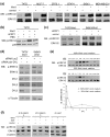
Autocrine WNT signaling in breast cancer cell lines. (a) Lysates from the indicated human breast cancer cell lines were analyzed by SDS-PAGE followed by immunoblotting for Dishevelled 1 (DVL1), DVL2, and DVL3 (the upper band indicates the phosphorylated form of each), active β-catenin, total β-catenin, and α-Tubulin as a loading control. (b) Lysates from T47D/Wnt1 cells and vector control were analyzed by SDS-PAGE/immunoblotting for active β-catenin, total β-catenin, and α-Tubulin. WB, Western blotting; WNT, wingless and integration site growth factor.

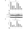
Treatment of human breast cancer cells with secreted Frizzled-related protein 1 (sFRP1) reduces proliferation and impairs canonical wingless and integration site growth factor signaling and extracellular signal-regulated kinase 1/2 (ERK1/2) phosphorylation. (a) One thousand to 5,000 cells of the indicated cell lines were seeded in a 96-well plate, and proliferation was measured in a YOPRO assay after 3 days of treatment with sFRP1 conditioned medium (CM) or control CM. (b) T47D cells were treated for 3 days with 30 μg/mL purified sFRP1 or phosphate-buffered saline, and cell numbers were measured in an MTT (3-[4,5-dimethylthiazol-2-yl]-2,5-diphenyl-tetrazolium bromide) assay. The results represent the mean of three experiments (± standard error). *p < 0.05, **p < 0.005, unpaired Student t test, comparison to corresponding control-treated cell line. (c) The indicated human breast cancer cell lines were treated for 2 hours with concentrated sFRP1 CM, and cell lysates were analyzed by SDS-PAGE/immunoblotting for active β-catenin, total β-catenin, p-ERK1/2, and ERK1/2 (upper panel). The results were quantified using ImageQuant (lower panel).

Short interfering RNA (siRNA)-mediated knockdown of Dishevelled (DVL) homologues results in decreased canonical wingless and integration site growth factor (WNT) signaling, a reduction in basal epidermal growth factor receptor (EGFR) and extracellular signal-regulated kinase 1/2 (ERK1/2) activation, and the induction of apoptosis in human breast cancer cells. (a) The indicated human breast cancer cell lines were transfected with pan-DVL siRNA. Two thousand to 5,000 cells were seeded in triplicate in 12-well plates the day after the transfection, and the cell number was counted after 7 days using a Vi-Cell XR cell viability analyzer. DVL knockdown was verified by SDS-PAGE/immunoblotting (only DVL3 is shown). The levels of act. β-catenin, total β-catenin, the WNT target c-MYC, and poly(ADP-ribose)polymerase (PARP) were analyzed by SDS-PAGE/immunoblotting. The lower band (80 kDa) in the blot probed for PARP represents the cleavage product upon induction of apoptosis. α-Tubulin was used as a loading control. For quantification, act. β-catenin levels were normalized to total β-catenin and c-MYC was normalized to α-Tubulin expression. (b) The indicated human breast cancer cell lines were transfected with pan-DVL siRNA and analyzed by SDS-PAGE/immunoblotting for p-ERK1/2 and EGFR Y845 phosphorylation. DVL2 levels are shown to monitor efficient knockdown of DVL, and α-Tubulin was used as loading control. For quantification, p-ERK1/2 was normalized to total ERK1/2 and p-EGFR Y845 was normalized to total EGFR expression.

Wnt1 induces rapid phosphorylation of extracellular signal-regulated kinase 1/2 (ERK1/2) in human breast cancer cells. (a) Cultures of the indicated cell lines were treated for 20 minutes with Wnt1 conditioned medium (CM) or control CM, and lysates were analyzed by SDS-PAGE followed by immunoblotting for p-ERK1/2 and ERK1/2. (b) T47D cells were treated with control CM or Wnt1 CM, which was previously incubated for 2 hours with 30 μg/mL purified secreted Frizzled-related protein 1 (sFRP1) or phosphate-buffered saline as control. Cell lysates were analyzed by SDS-PAGE/immunoblotting for p-ERK1/2 and p38 as loading control. (c) Stably Wnt1-transfected T47D and SkBr3 cells were treated for 2 hours with sFRP1 CM, control CM, or normal growth medium. Total lysates were analyzed by SDS-PAGE followed by immunoblotting for p-ERK1/2 and ERK1/2. (d) T47D/Wnt1 and SkBr3/Wnt1 cells were seeded at 300,000 cells per well in a six-well plate the day before short interfering RNA (siRNA) transfection with either a LacZ control siRNA or pan-DVL siRNA. The cells were cultured for an additional 48 hours under normal growth conditions and 24 hours in 0.1% fetal calf serum before harvesting. Total lysates were analyzed by SDS-PAGE/immunoblotting for p-ERK1/2, ERK1/2, DVL1, DVL2, and DVL3, or α-Tubulin as a loading control. (e) T47D cultures were treated for the indicated times with 100% and 20% vol/vol Wnt1 CM. Total lysates were analyzed by SDS-PAGE/immunoblotting for p-ERK1/2 and ERK1/2 (upper panel). ERK activation was quantified after normalization of signal intensity of p-ERK1/2 to total ERK1/2 using the ImageQuant software. ERK activity peaks at between 30 minutes and 1 hour (lower panel). (f) Wnt1-mediated effects are independent of β-catenin. T47D cells were infected with a retrovirus carrying an expression cassette for a short-hairpin RNA targeting β-catenin. Two independent shβ-catenin clones and a control pool infected with a short hairpin against bacterial LacZ were treated with control CM or Wnt1 CM for 20 minutes or 10 ng/mL EGF for 5 minutes. Total lysates were analyzed by SDS-PAGE/immunoblotting for p-ERK1/2, ERK1/2, and β-catenin. DVL, Dishevelled.

Wnt1-induced extracellular signal-regulated kinase 1/2 (ERK1/2) phosphorylation depends on epidermal growth factor receptor (EGFR) activity. (a) EGFR was immunoprecipitated from 2 mg of whole cell lysate from T47D/Wnt1, T47D/vector, SkBr3/Wnt1, and SkBr3 vector cells and analyzed by SDS-PAGE/immunoblotting for p-Tyr and EGFR. The p-Tyr signal corresponding to EGFR was quantified using the ImageQuant software. (b) T47D/Wnt1 cells were pre-treated for 1 hour with monoclonal antibody 225 containing conditioned medium (CM), and lysates were analyzed by SDS-PAGE/immunoblotting for p-ERK1/2 and ERK1/2. (c) T47D/Wnt1 cells were pre-treated for 1 hour with the metalloprotease inhibitor CGS27023A (50 μM) or dimethyl sulfoxide as control. Total lysates were analyzed by SDS-PAGE/immunoblotting for p-ERK1/2 and ERK1/2. (d) T47D cells were pre-treated for 1 hour with 2 μM PKI166 before treatment with Wnt1 CM or control CM. T47D/Wnt1 and SkBr3/Wnt1 cells were treated for 1 hour with 2 μM PKI166 prior to lysing the cells. Lysates were analyzed by SDS-PAGE/immunoblotting for p-ERK1/2 and ERK1/2. Tyr, tyrosine.

Src kinase is required for Wnt1-mediated extracellular signal-regulated kinase 1/2 (ERK1/2) activation. (a) c-Src was immunoprecipitated from lysates of Wnt1-expressing or vector control-expressing SkBr3 cells. The immunoprecipates were analyzed by SDS-PAGE/immunoblotting for p-Src and Src, and the signals were quantified versus control levels using the ImageQuant program. (b) T47D cells were pre-treated with the Src kinase inhibitor CGP77675 (2 μM) or dimethyl sulfoxide (DMSO) for 1 hour before treatment for 20 minutes with Wnt1 conditioned medium (CM). T47D/Wnt1 and SkBr3/Wnt1 cells were treated for 1 hour with CGP77675 or DMSO. Total lysates were analyzed by SDS-PAGE/immunoblotting for p-ERK1/2 and ERK1/2. (c) Src-/- mouse embryonic fibroblasts transfected with a control plasmid or a wild type Src-expressing vector were treated with Wnt1 CM or control CM for 20 minutes. Total lysates were analyzed by SDS-PAGE/immunoblotting for p-ERK1/2 and ERK1/2. A representative result of three independent experiments is shown. EGF, epidermal growth factor; IP, immunoprecipitation; WB, Western blotting.

Wnt1 rescues breast tumor cells from the anti-proliferative effects of 4-hydroxytamoxifen (4-HT). T47D (a) or MCF-7 (b) cells were treated with 4-HT (5 μM), PKI166 (5 μM), or a combination of both drugs for 7 or 6 days, respectively, in the presence or absence of Wnt1 conditioned medium (CM) or control CM. For control experiments, the solvents ethanol and dimethyl sulfoxide, respectively, were added in corresponding concentrations. Cultures were re-fed with fresh medium and inhibitors after 3 days, before cell number (± standard error) was determined. For biochemical analysis, T47D cells were treated under corresponding conditions with 4-HT (5 μM), AEE788 (2 μM), or a combination of both in the absence or presence of Wnt1 CM or control CM for 2 hours or 3 days. Total lysates were analyzed by SDS-PAGE/immunoblotting for p-ERK1/2 and ERK1/2. *p < 0.05, **p < 0.005, unpaired Student t test. ERK1/2, extracellular signal-regulated kinase 1/2; n.s., difference not significant; WB, Western blotting.

Schematic representation of wingless and integration site growth factor (WNT)-induced epidermal growth factor receptor (EGFR) transactivation. Our results show that Wnt1 induces a signaling cascade that links the activation of EGFR in a manner dependent on Dishevelled, SRC, metalloprotease, and EGF-like ligand to the sFRP1-sensitive activation of Frizzled receptors. The activation of EGFR, which may occur via phosphorylation of Y845, an SRC phosphorylation site, triggers activation of the extracellular signal-regulated kinase 1/2 (ERK1/2) pathway. Together with the canonical WNT/β-catenin pathway and its target genes, including c-Myc, the ERK1/2 pathway promotes proliferation and survival of breast cancer cells. Furthermore, activation of the ERK1/2 signaling by Wnt1 may contribute to the development of anti-estrogen resistance. DVL, Dishevelled; EGF, epidermal growth factor; ER, estrogen receptor; sFRP1, secreted Frizzled-related protein 1.
Similar articles
-
Matsuda Y, Schlange T, Oakeley EJ, Boulay A, Hynes NE. Matsuda Y, et al. Breast Cancer Res. 2009;11(3):R32. doi: 10.1186/bcr2317. Epub 2009 May 27. Breast Cancer Res. 2009. PMID: 19473496 Free PMC article.
-
Dishevelled: The hub of Wnt signaling.
Gao C, Chen YG. Gao C, et al. Cell Signal. 2010 May;22(5):717-27. doi: 10.1016/j.cellsig.2009.11.021. Epub 2009 Dec 13. Cell Signal. 2010. PMID: 20006983 Review.
-
A Wnt-ow of opportunity: targeting the Wnt/beta-catenin pathway in breast cancer.
Prosperi JR, Goss KH. Prosperi JR, et al. Curr Drug Targets. 2010 Sep;11(9):1074-88. doi: 10.2174/138945010792006780. Curr Drug Targets. 2010. PMID: 20545611 Review.
Cited by
-
Multiplexed protein profiling reveals spatial subcellular signaling networks.
Cai S, Hu T, Venkatesan M, Allam M, Schneider F, Ramalingam SS, Sun SY, Coskun AF. Cai S, et al. iScience. 2022 Aug 18;25(9):104980. doi: 10.1016/j.isci.2022.104980. eCollection 2022 Sep 16. iScience. 2022. PMID: 36093051 Free PMC article.
-
Cooper SJ, von Roemeling CA, Kang KH, Marlow LA, Grebe SK, Menefee ME, Tun HW, Colon-Otero G, Perez EA, Copland JA. Cooper SJ, et al. Mol Cancer Ther. 2012 Oct;11(10):2105-15. doi: 10.1158/1535-7163.MCT-11-0873. Epub 2012 Jul 23. Mol Cancer Ther. 2012. PMID: 22826467 Free PMC article.
-
Hawkins S, Namboori SC, Tariq A, Blaker C, Flaxman C, Dey NS, Henley P, Randall A, Rosa A, Stanton LW, Bhinge A. Hawkins S, et al. Stem Cell Reports. 2022 Jul 12;17(7):1650-1665. doi: 10.1016/j.stemcr.2022.05.019. Epub 2022 Jun 23. Stem Cell Reports. 2022. PMID: 35750046 Free PMC article.
-
Gracanin A, Timmermans-Sprang EP, van Wolferen ME, Rao NA, Grizelj J, Vince S, Hellmen E, Mol JA. Gracanin A, et al. PLoS One. 2014 Jun 2;9(6):e98698. doi: 10.1371/journal.pone.0098698. eCollection 2014. PLoS One. 2014. PMID: 24887235 Free PMC article.
-
Chen Q, Cao HZ, Zheng PS. Chen Q, et al. Oncotarget. 2014 Oct 15;5(19):9092-105. doi: 10.18632/oncotarget.2377. Oncotarget. 2014. PMID: 25193857 Free PMC article.
References
Publication types
MeSH terms
Substances
LinkOut - more resources
Full Text Sources
Other Literature Sources
Medical
Research Materials
Miscellaneous