Cutaneous Mycobacterial Infections - PubMed
- ️Mon Jan 01 2018
Review
. 2018 Nov 14;32(1):e00069-18.
doi: 10.1128/CMR.00069-18. Print 2018 Jan.
Affiliations
- PMID: 30429139
- PMCID: PMC6302357
- DOI: 10.1128/CMR.00069-18
Review
Cutaneous Mycobacterial Infections
Carlos Franco-Paredes et al. Clin Microbiol Rev. 2018.
Abstract
Humans encounter mycobacterial species due to their ubiquity in different environmental niches. In many individuals, pathogenic mycobacterial species may breach our first-line barrier defenses of the innate immune system and modulate the activation of phagocytes to cause disease of the respiratory tract or the skin and soft tissues, sometimes resulting in disseminated infection. Cutaneous mycobacterial infections may cause a wide range of clinical manifestations, which are divided into four main disease categories: (i) cutaneous manifestations of Mycobacterium tuberculosis infection, (ii) Buruli ulcer caused by Mycobacterium ulcerans and other related slowly growing mycobacteria, (iii) leprosy caused by Mycobacterium leprae and Mycobacterium lepromatosis, and (iv) cutaneous infections caused by rapidly growing mycobacteria. Clinically, cutaneous mycobacterial infections present with widely different clinical presentations, including cellulitis, nonhealing ulcers, subacute or chronic nodular lesions, abscesses, superficial lymphadenitis, verrucous lesions, and other types of findings. Mycobacterial infections of the skin and subcutaneous tissue are associated with important stigma, deformity, and disability. Geography-based environmental exposures influence the epidemiology of cutaneous mycobacterial infections. Cutaneous tuberculosis exhibits different clinical phenotypes acquired through different routes, including via extrinsic inoculation of the tuberculous bacilli and dissemination to the skin from other sites, or represents hypersensitivity reactions to M. tuberculosis infection. In many settings, leprosy remains an important cause of neurological impairment, deformity, limb loss, and stigma. Mycobacterium lepromatosis, a mycobacterial species related to M. leprae, is linked to diffuse lepromatous leprosy of Lucio and Latapí. Mycobacterium ulcerans produces a mycolactone toxin that leads to subcutaneous tissue destruction and immunosuppression, resulting in deep ulcerations that often produce substantial disfigurement and disability. Mycobacterium marinum, a close relative of M. ulcerans, is an important cause of cutaneous sporotrichoid nodular lymphangitic lesions. Among patients with advanced immunosuppression, Mycobacterium kansasii, the Mycobacterium avium-intracellulare complex, and Mycobacterium haemophilum may cause cutaneous or disseminated disease. Rapidly growing mycobacteria, including the Mycobacterium abscessus group, Mycobacterium chelonei, and Mycobacterium fortuitum, are increasingly recognized pathogens in cutaneous infections associated particularly with plastic surgery and cosmetic procedures. Skin biopsies of cutaneous lesions to identify acid-fast staining bacilli and cultures represent the cornerstone of diagnosis. Additionally, histopathological evaluation of skin biopsy specimens may be useful in identifying leprosy, Buruli ulcer, and cutaneous tuberculosis. Molecular assays are useful in some cases. The treatment for cutaneous mycobacterial infections depends on the specific pathogen and therefore requires a careful consideration of antimicrobial choices based on official treatment guidelines.
Keywords: Buruli ulcer; Mycobacterium; Mycobacterium kansasii; Mycobacterium marinum; Mycobacterium ulcerans; cutaneous; leprosy; mycobacteria; nontuberculous mycobacteria; tuberculosis.
Copyright © 2018 American Society for Microbiology.
Figures

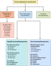
Classification of major pathogenic mycobacteria.

Tuberculosis verrucosa cutis of the hand, manifesting as verrucous plaques caused by direct inoculation of the tuberculous bacilli into the skin of an individual previously sensitized to this pathogen.

Scrofuloderma presenting in the neck, resulting from direct extension of an infected left cervical lymph node into the overlying cutaneous structures. This form is also known as tuberculosis colliquative cutis. This form of cutaneous tuberculosis is also associated with infection caused by Mycobacterium bovis or bacillus Calmette-Guérin.

Clinical manifestations of leprosy: borderline tuberculoid (BT) (A), borderline borderline (BB) (B), and lepromatous (LL) (C).

An 11-year-old male demonstrating a destructive panniculitis causing ulceration with undermined borders, characteristic of Buruli ulcer.

An adult with Mycobacterium abscessus infection presenting as scrofuloderma with extensive tissue destruction in the right cervical and supraclavicular areas.

Infection caused by Mycobacterium fortuitum associated with mesotherapy.

Characteristic sporotrichoid nodular lymphangitic spread of Mycobacterium marinum.

Severe hand swelling and nodular lymphangitic lesions caused by Mycobacterium marinum infection.

Mycobacterium kansasii leading to a sporotrichoid nodular lymphangitis of the right arm.

Cold abscess caused by Mycobacterium avium-intracellulare complex infection in a 60-year-old male.
Similar articles
-
Overview of Cutaneous Mycobacterial Infections.
Franco-Paredes C, Chastain DB, Allen L, Henao-Martínez AF. Franco-Paredes C, et al. Curr Trop Med Rep. 2018 Dec;5(4):228-232. doi: 10.1007/s40475-018-0161-7. Epub 2018 Aug 3. Curr Trop Med Rep. 2018. PMID: 34164254 Free PMC article.
-
[Cutaneous and soft skin infections due to non-tuberculous mycobacteria].
Alcaide F, Esteban J. Alcaide F, et al. Enferm Infecc Microbiol Clin. 2010 Jan;28 Suppl 1:46-50. doi: 10.1016/S0213-005X(10)70008-2. Enferm Infecc Microbiol Clin. 2010. PMID: 20172423 Review. Spanish.
-
Recent advances in leprosy and Buruli ulcer (Mycobacterium ulcerans infection).
Walsh DS, Portaels F, Meyers WM. Walsh DS, et al. Curr Opin Infect Dis. 2010 Oct;23(5):445-55. doi: 10.1097/QCO.0b013e32833c2209. Curr Opin Infect Dis. 2010. PMID: 20581668 Review.
-
Identification of mycobacterial DNA in cutaneous lesions of sarcoidosis.
Li N, Bajoghli A, Kubba A, Bhawan J. Li N, et al. J Cutan Pathol. 1999 Jul;26(6):271-8. doi: 10.1111/j.1600-0560.1999.tb01844.x. J Cutan Pathol. 1999. PMID: 10472755
-
Nontuberculous Mycobacteria: Skin and Soft Tissue Infections.
Gonzalez-Santiago TM, Drage LA. Gonzalez-Santiago TM, et al. Dermatol Clin. 2015 Jul;33(3):563-77. doi: 10.1016/j.det.2015.03.017. Epub 2015 May 8. Dermatol Clin. 2015. PMID: 26143432 Review.
Cited by
-
López-Roa P, Esteban J, Muñoz-Egea MC. López-Roa P, et al. Microorganisms. 2022 Dec 29;11(1):90. doi: 10.3390/microorganisms11010090. Microorganisms. 2022. PMID: 36677382 Free PMC article. Review.
-
Eyer-Silva WA, Almeida MR, Martins CJ, Basílio-de-Oliveira RP, Araujo LF, Basílio-de-Oliveira CA, Azevedo MCVM, Pinto JFDC, Vasconcellos SEG, Rodrigues-Dos-Santos Í, MagdinierGomes H, Suffys PN. Eyer-Silva WA, et al. Rev Inst Med Trop Sao Paulo. 2019 Dec 20;61:e71. doi: 10.1590/S1678-9946201961071. eCollection 2019. Rev Inst Med Trop Sao Paulo. 2019. PMID: 31859848 Free PMC article.
-
Ueda Y, Okamoto T, Sato Y, Hayashi A, Takahashi T, Kamada K, Honda S, Hotta K. Ueda Y, et al. J Nephrol. 2022 Sep;35(7):1907-1910. doi: 10.1007/s40620-021-01244-2. Epub 2022 Jan 4. J Nephrol. 2022. PMID: 34982412 No abstract available.
-
Genomic characteristics of Mycobacterium tuberculosis isolates of cutaneous tuberculosis.
Mei YM, Zhang WY, Sun JY, Jiang HQ, Shi Y, Xiong JS, Wang L, Chen YQ, Long SY, Pan C, Luo T, Wang HS. Mei YM, et al. Front Microbiol. 2023 May 17;14:1165916. doi: 10.3389/fmicb.2023.1165916. eCollection 2023. Front Microbiol. 2023. PMID: 37266022 Free PMC article.
-
Maimaiti Z, Li Z, Xu C, Fu J, Hao L, Chen J, Li X, Chai W. Maimaiti Z, et al. Orthop Surg. 2023 Jun;15(6):1488-1497. doi: 10.1111/os.13661. Epub 2023 May 8. Orthop Surg. 2023. PMID: 37154097 Free PMC article. Review.
References
Publication types
MeSH terms
LinkOut - more resources
Full Text Sources
Other Literature Sources
Medical